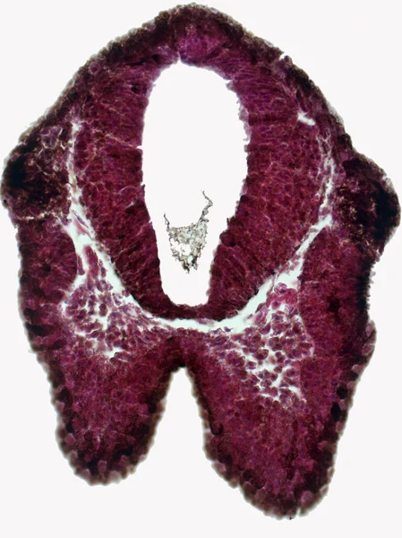
Eye Development Cells Both Mesodermal Ectodermal Tissues Contribute Formation Eye — 图库照片

Eye development. Cells from both the mesodermal and the ectodermal tissues contribute to the formation of the eye. In the center is a crystalline lens. — 图库图片

Eye development. Cells from both the mesodermal and the ectodermal tissues contribute to the formation of the eye. In the center is a crystalline lens.

 — 照片作者 BioFoto

同一系列:

Cross section of the olfactory placode and brain of the embryo frog. Hematoxylin and Eosin Staining (H&E stain).
Cross Section Olfactory Placode Brain Embryo Frog Hematoxylin Eosin Staining — 图库照片
伪极性神经元。背根神经节的圆体细胞瘤的假非极性神经元被称为卫星细胞的胶质细胞所包围。血清素和血清素染色。放大率:x1000.
伪极性神经元 背根神经节的圆体细胞瘤的假非极性神经元被称为卫星细胞的胶质细胞所包围 血清素和血清素染色 放大率 X1000 — 图库照片
小脑的横断面轻缩微图.
小脑的横断面轻缩微图 — 图库照片
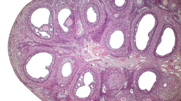
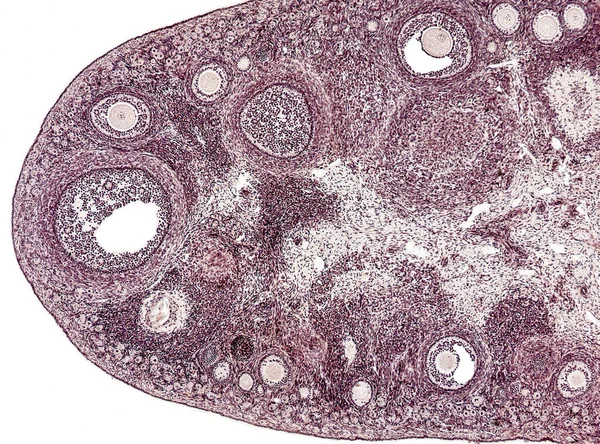
Light micrograph of the Cat's ovarian. Micrograph of ovary showing primordial, primary and secondary follicles isolated on white background. Hematoxylin and eosin stain.
Light Micrograph Cat Ovarian Micrograph Ovary Showing Primordial Primary Secondary — 图库照片
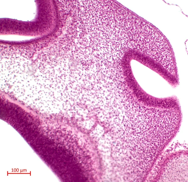
Olfactory organ development. Olfactory fossa pit stage. Hematoxylin and Eosin Staining (H&E stain).
Olfactory Organ Development Olfactory Fossa Pit Stage Hematoxylin Eosin Staining — 图库照片
雄松(Pinus sylvestris)锥的纵向切片,显示小孢子和花粉.
Pinus Sylvestris 锥的纵向切片 显示小孢子和花粉 — 图库照片
小脑的横断面轻缩微图.
小脑的横断面轻缩微图 — 图库照片
骨骼肌横断面上的条带肌纤维显示,在细胞周围有几个多核细胞。光镜摄影显微镜.
骨骼肌横断面上的条带肌纤维显示 在细胞周围有几个多核细胞 光镜摄影显微镜 — 图库照片
简单的柱状上皮。肾髓质的光镜,显示由一个简单的柱状上皮细胞内衬的几个收集导管。放大率:1 000
简单的柱状上皮 肾髓质的光镜 显示由一个简单的柱状上皮细胞内衬的几个收集导管 放大率 000 — 图库照片
用显微镜拍的一张有趣的照片周围神经中的非骨髓化纤维。纵向部分。血红素和血红素.
用显微镜拍的一张有趣的照片周围神经中的非骨髓化纤维 纵向部分 血红素和血红素 — 图库照片
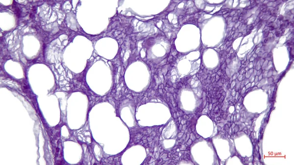
光镜:管状骨的白色脂肪组织.大而明亮的孔-脂肪细胞的残余.
管状骨的白色脂肪组织 大而明亮的孔 脂肪细胞的残余 — 图库照片
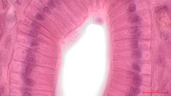
通过气管(或气管)的部分的光镜,气管是从口通往支气管和肺的管子。气管腔内衬有呼吸上皮细胞.
通过气管 或气管 的部分的光镜 气管是从口通往支气管和肺的管子 气管腔内衬有呼吸上皮细胞 — 图库照片
人体肾组织,光镜。Haematoxylin和Eosin染色。肾小管的立方体上皮细胞由一层立方体细胞组成.
人体肾组织 Haematoxylin和Eosin染色 肾小管的立方体上皮细胞由一层立方体细胞组成 — 图库照片
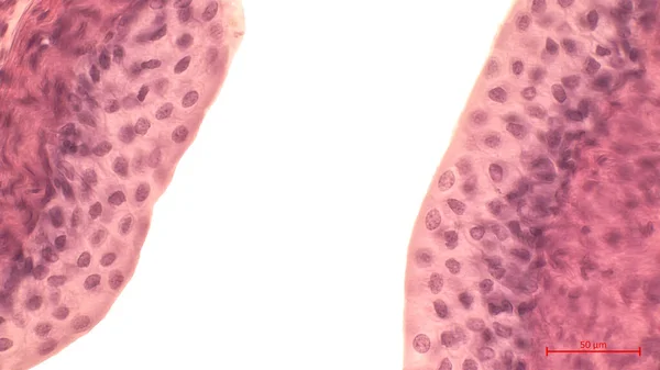
膀胱内衬的光镜检查.该组织是一个广泛的过渡上皮细胞(urothelium),因为它能适应膀胱大小的变化.
膀胱内衬的光镜检查 该组织是一个广泛的过渡上皮细胞 Urothelium 因为它能适应膀胱大小的变化 — 图库照片
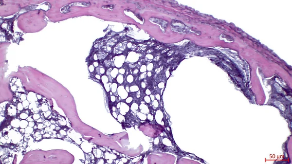
骨头横断面的光镜。一种发育中的长骨的窒息.
骨头横断面的光镜 一种发育中的长骨的窒息 — 图库照片
小脑的横断面轻缩微图.
小脑的横断面轻缩微图 — 图库照片

使用信息

您可以根据标准或扩展许可将此免版税图片"Eye development. Cells from both the mesodermal and the ectodermal tissues contribute to the formation of the eye. In the center is a crystalline lens."用于个人和商业目的。标准许可涵盖了大多数用途,包括广告、UI 设计和产品包装,允许多达 500,000 份打印副本。扩展许可允许标准许可下的所有用途,带无限制的打印权限,并允许您将下载的库存图片用于商品、产品转售或免费分发。

您可以购买此库存图片,并以高达5600x3207的高分辨率下载。 上传日期: 2022年5月15日